Breast Cancer Image Classification
Implementing various Neural Network architectures to perform Breast Cancer Image Classification
1. Introduction
Breast cancer remains a global health issue, necessitating innovative approaches to enhance early detection and diagnosis. Histopathological examination of tissue samples is a crucial diagnostic technique, providing valuable insights into the nature of tumors.
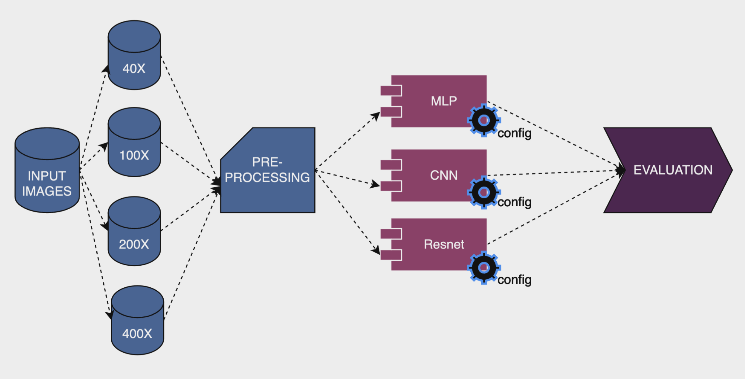
This research aims to implement various Neural Network architectures, including Multi-Layer Perceptron (MLP), Convolutional Neural Networks (CNN), and Residual Networks (ResNet), to perform breast cancer histopathological images classification.
2. Dataset
The dataset includes both benign (non-cancerous) and malignant (cancerous) cases captured at varying magnification levels (40x, 100x, 200x, and 400x), from The Breast Cancer Histopathological Database (BreakHis)
3. Method
The main goal is to compare the performances of various architectures within three different models: MLP, CNN, and ResNet, to determine the most robust and performant architecture.

The BreakHis dataset, which includes breast cancer histopathological images at various magnification levels, is used for the experiments. Then, I train the models, which I re-implement from scratch, on the Training set and fine-tune them with diverse hyperparameter values using the Validation set. Finally, best models are selected based on their average accuracy across four magnifications on the Validation set and report their final average accuracy over these magnifications on the Test set.
4. Usage
- You can find the source code of the project here